Background
Whole slide imaging information is potentially available for virtually all cancer
patients as Pathology studies are, with very few exceptions, carried out for cancer
patients. Information extracted from digitized pathology images (Pathomics data) can
increasingly be employed to generate precise characterizations of a patient’s cancer
– identification and classification of cells and characterization of tumor microenvironment.
This information can be used, in the context of clinical, molecular and Radiology
information to create imaging biomarkers used to predict outcome and steer treatment;
this information can also be employed to clinical decision support systems to improve
reproducibility and precision of traditional Pathology reports. Continue Reading...
|
The IEDM will develop and provide the methods and tools to integrate and analyze detailed
morphology and spatially mapped molecular data and to allow researchers to gain crucial
insights into their scientific problems with the aid, and further development, of
engineering tools including artificial intelligence and machine learning.
The need for deep, quantitative understanding of biomedical systems is crucial to
unravel the underlying mechanisms and pathophysiology. Technologies developed by the
IEDM, particularly this thrust, will enable researchers to assemble and visualize
detailed, multi-scale descriptions of tissue morphologic changes originating from
a wide range of microscopy instruments and provide the computational and pattern recognition
tools to integrate these descriptions with corresponding genomic, proteomic, glycemic
and clinical signatures. Together these capabilities will facilitate and propel research
and discovery in a range of pivotal, cutting-edge biomedical projects with the aid
of the latest engineering advances in digital technology including machine learning
and artificial intelligence.
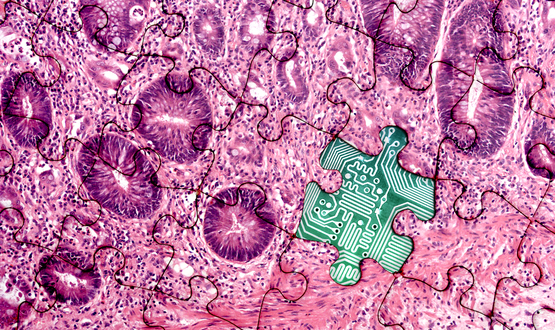
Pathomics, or the automated quantification of a pathology image-based phenotype, is
increasingly seen as a key enabler for precision medicine. Over the past 20 years
digital pathology has developed into a rapidly growing field with applications in
translational research. Pathomics analyses characterize cells and tissue obtained
in pathology studies. The results of a pathomics study is fundamentally different
from a pathologist’s report. A pathologist report describes what the pathologist observes
when inspecting tissue, while pathomics features provide a quantitative and reproducible
characterization of that tissue. Examples of pathomics features include: 1) spatial
characterization of tumor and stroma regions, 2) shapes and textures of nuclei, 3)
classifications of cell type, and 4) quantitative characterization of lymphocytic
infiltration. Pathomics analyses have been shown to provide value in a variety of
correlative and prognostic studies.
Quantitative characterization of tumor infiltrating lymphocytes (TILs), for example,
is of rapidly increasing importance in precision medicine. With the growth of cancer
immunotherapy, these characterizations are likely to be of increasing clinical significance,
as understanding each patient’s immune response becomes more important. High densities
of TILs correlate with favorable clinical outcomes including longer disease-free survival
or improved overall survival (OS) in multiple cancer types. Recent studies further
suggest that the spatial context and the nature of cellular heterogeneity within the
tumor microenvironment, in terms of the immune infiltrate into the tumor center and
invasive margin, are important in cancer prognosis.
Prognostic factors, most notably the immunoscore, that quantify spatial TIL densities
in different tumor regions, have high prognostic value. Hence, assessments of tumor-associated
lymphocytes are increasingly important both in the clinical assessment of pathology
slides, and in translational research into the role of these lymphocytic populations.
Faculty and other researchers in the Stony Brook Departments of Pathology and Medicine
have conducted leading research in understanding cancer molecular biology for pancreatic
cancer, urothelial and prostate cancer, breast cancer, and leukemia, as well as colorectal cancer. They are developing diagnostic biomarkers and therapeutic targets, and potentially
intervening in these processes through drug discovery.
|
Faculty Contributors
Euvgenia Alexandrova, Pathology
Eric Brouzes, Biomedical Engineering
Chao Chen, Computer Science
Jun Chung, Pathology
Geoff Girnun, Pathology
John Haley, Pathology
Jingfang Ju, Pathology
Richard Kew, Pathology
Tahsin Kurc, Biomedical Informatics
Yupo Ma, Pathology
Natalia Marchenko, Pathology
Luis Martinez, Pathology
David Matus, Biochemistry & Cell Biology
|
|
Richard Moffitt, Biomedical Informatics
David Montrose, Pathology
Scott Powers, Pathology
Joel Saltz, Biomedical Informatics
Dimitris Samaras, Computer Science
I.V. Ramakrishnan, Computer Science
Kanokporn Rithidech, Pathology
Adam Rosebrock, Pathology
Kenneth Shroyer, Pathology
Flaminia Talos, Pathology/Urology
Patricia Thompson, Pathology
Fusheng Wang, Computer Science
|
|