Machine Learning Diagnosis of In Vivo Tissues by Extracting Tissue-Energy Characteristic Features from Non-Invasively Acquired Computed Tomographic Data
Gorodenkoff, https://stock.adobe.com/uk/226212015, stock.adobe.com
Background
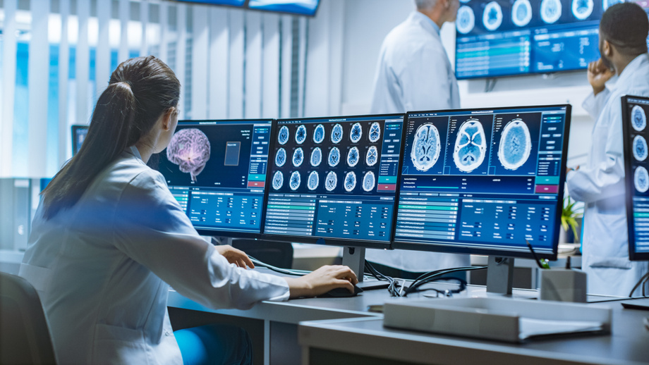
Computed tomography (CT) is a widely used imaging tool for early detection of cancers such as lung and colorectal cancer. However, current low-dose CT screening protocols suffer from high false positive rates, particularly in the classification of indeterminate lesions (IDLs), which often lead to costly and invasive follow-up procedures. This challenge stems in part from the way CT images are generated—averaging tissue responses over a broad X-ray energy spectrum— resulting in loss of diagnostic detail. Existing machine learning (ML) approaches have not substantially improved diagnostic accuracy, in part because they overlook critical prior knowledge about how different tissues uniquely respond to varying X-ray energies. There remains a need for more precise, non-invasive diagnostic tools that can reduce false positives (FPs) and better distinguish malignant from benign lesions in CT screening data.
Technology
This technology decomposes poly-energetic CT images into mono-energetic tissue distribution maps (MTMs) and extracts tissue elasticity measures at each energy level to create tissue-energy characteristic features. These features enable ML models to more accurately classify indeterminate pulmonary nodules and colorectal polyps from low-dose CT screening data, surpassing current diagnostic methods by significantly improving the area under the receiver operating characteristic curve (AUC) from the 0.60-0.80 range to the 0.90s. This approach addresses the limitations of traditional CT imaging that averages tissue responses across a wide X- ray energy spectrum, thereby enhancing early cancer detection and reducing FPs in lung and colorectal cancer screening.
Advantages
Incorporates prior knowledge of tissue-specific responses to different X-ray energies for improved feature extraction. - Generates mono-energetic tissue distribution maps or MTMs to capture energy-sensitive tissue characteristics. - Utilizes tissue elasticity measures as biological discriminative features for classification of lesion malignancy. - Eliminates redundancy while integrating the biological discriminative features from different individual energy levels to achieve the highest AUC. - Significantly boosts diagnostic accuracy with AUC improvements into the 0.90s for IDLs. - Reduces false positive rates in low-dose CT cancer screening, potentially lowering unnecessary follow-up procedures. - Applicable to challenging indeterminate lesions that are difficult to diagnose using conventional methods.
Application
Early detection and diagnosis of lung cancer through improved low-dose CT screening. - Colorectal cancer screening with enhanced lesion malignancy prediction. - Medical imaging diagnostic tools integrating machine learning for cancer lesion classification. - Healthcare providers aiming to reduce false positives and improve patient management in cancer screening programs. - Development of advanced AI-driven radiology software solutions.
Inventors
Jerome Liang, Professor, Radiology
Licensing Potential
Development partner - Commercial partner - Licensing
Licensing Status
Available for licensing
Licensing Contact
Valery Matthys, Licensing Associate, Intellectual Property Partners, valery.matthys@stonybrook.edu,
Patent Status
Patent application submitted.
Stage of Development
In Vivo
Tech ID
050-9396