Journal
19 July 2011
Finally finding a set of labeled neutral density filters, I was able to begin taking data, or so I thought. The DMM that I found the previous day unfortunately only read current in mA. What I found upon turning on the laser was that I needed to take my readings in μA. Luckily, I noticed that we had a variety of DMM's that were able to measure current at this order of magnitude. Unfortunately, none of the batteries could hold up for more than a minute without dying. I finally was able to find a sufficient 9V battery and was able to continue my project without having the meters shut down every minute or so.
I continued to take data for the entire day, excluding the time in which I attended the weekly REU seminar. This week's seminar was a presentation on high energy particle physics by Dr. Hemmick. The talk was interesting and Dr. Hemmick was very enthusiastic about the topic. If I had had a bit more experience with the topic, I would have understood it better than I did. Overall, it introduced me to an exciting area of physics research that I did not have much knowledge of previously!
18 July 2011
Today I spent time working on diagrams for my project page and read more information concerning beam profiling while the laser was used in the materials science building. When I went to pick up the laser, I worked with Dr. Sokolov and a man from the engineering machine shop to devise a mount for the laser and associated optical elements. We settled upon attaching a heat sink to the laser and mounting that upon a post/holder system. This will then be attached to a metal arm that can be shifted left and right as well as upward and downward. The metal arm will have evenly spaced holes upon which other optical elements can be assembled to enhance the effectiveness of the laser in fluorescing the DNA samples. I am slightly worried about the assembly because I believe that the distance in which the optical elements will be placed from the laser will severely limit the range of angles at which the laser can be used to illuminate the specimen. Also, I noticed that the laser, when illuminating at an angle almost parallel to a test specimen (filter paper), created an elliptical spot. The spot was very exaggerated. I do not know whether this will negatively impact the fluorescence of the DNA samples. If so, we may have to distinguish a minimum angle at which successful illumination can be achieved.
After designing its mount, I brought the laser back to the LTC to work on determining the profile of its beam. I set up the laser with a photodetector and a razor blade on a translation stage as discussed in the literature that I was reading. Before turning on the laser, I worked with a power supply to make sure that I got a feel for the sensitivity of the voltage knobs. This way, I will not accidentally push the laser passed its maximum voltage input when I turn it on. To be safe, we have not been running the laser at its maximum input voltage. Since we have been doing most of the measurements at a maximum of 2V, I decided to profile the beam at this input voltage. To make sure that I did not harm the photodetector, I looked for a neutral density filter to place in the optical train. I did not find any that were labeled, so I spent the rest of the day looking for better options.
14 July 2011 - 15 July 2011
Finding out about issues with the polarization orientation and extinction ratio associated with the laser from Dr. Sokolov, I decided that I needed to read through literature concerning laser polarization in hopes of finding explanations for the problems we were encountering as well as possible solutions to them. Unfortunately after two days of research, I could not find any explanations supporting the fact that our laser exhibited extreme extinction ratio dropoff at decreasing voltages. Since I could not find any literature on the problem, I could not find any solutions.
In addition to troubleshooting polarization issues, I needed to figure out some way to profile the beam put out by the laser. I found that there are beam profilers available to do this, but they are expensive and I am sure that we do not have one. As I was reading, I also learned about different "width" terminology when discussing the profile of a laser beam including D4&#sigma;, knife edge, D86 and 1/e**2. I believe that the knife edge technique will be the easiest to experiment with, but I still need to read more about it before I attempt it to make sure that I do not damage the laser or photodetector.
13 July 2011
At the LTC lunch seminar, I presented my experimentation with the DPSS laser and the reason behind attempting to use it in an oblique fashion. I also posed specific questions to the mentors in hopes of gaining some insight into different current problems concerning polarization determination and experiment results as well as to troubleshoot future problems we may encounter. I was much more confident in my presentation today and tried to relax as I explained the experimentation at hand. Hopefully with the mentors now understanding the point of my work, I will be able to go to them for assistance as the project moves on.12 July 2011
I spent the majority of the day in Dr. Sokolov's lab in the engineering building working on the DPSS. We effectively mounted the laser and set it up on a small breadboard. We then attempted to determine the direction of polarization of the DPSS beam using a Glan-Thompson polarizer on a rotational mount. We agree that the most likely orientation of the polarization is vertical. Using this information, we attempted to rotate the polarization of the beam using a half wave plate. I had found two in the lab the previous day that may have had a chance of working on the 473 nm laser, so we experimented with both plates. Only one of the plates was able to give decent results, but was still only able to work with 60% efficiency. I was also concerned that the photodetector was being saturated by the DPSS beam and that in future experimentation, we should use a neutral density filter to eliminate this variable.
11 July 2011
Today I met with Dr. Sokolov to discuss the laser project and meet the two high school students that would be working on its biological aspects. Prior to the 5:00 meeting, I scoured the lab for half wave plates that could possibly work correctly when placed in front of a 473 nm wavelength laser beam. I tested all of the wave plates that I found on the Argon laser in the back room. Because the Argon laser beam could be of any wavelength ranging from 457-514 nm (as notated on the product itself) I assumed that any wave plate that worked on the Argon laser could possibly have a chance of working on the DPSS. Hopefully one of the plates that I found will work to a somewhat efficient degree so that we may use it to rotate the polarization of the DPSS.
During my meeting with Dr. Sokolov, we reviewed the overall idea of the project and discussed the various aspects of the laser that would need to be tested before designing an optical system to send the beam through. I spent the rest of the night learning/reviewing polarization so that I would better understand the experiments planned for the next day.
8 July 2011
Today the other REU members and I took a trip to the American Museum of Natural History in New York City. I had just recently been to the museum in the spring, but was exited to be able to visit again. The museum is of incredible magnitude and there is always something new and interesting to learn.
In my previous visit, I simply walked around the museum and attended the planetarium show. This time around, the group and I attended more of the special exhibits which were extremely interesting! We went to the planetarium show that I had seen previously, but also walked through the "Brain" and "World's Largest Dinosaurs" exhibits. Having an attraction to biology, I found the "Brain" exhibit to be fascinating. It described neural connections and the functions of certain areas of the brain. The exhibit also showed how the brain controls voluntary and involuntary actions and how humans are able to learn. It was an incredible exhibit; I wish I could have spent more time in it!
After visiting the exhibits and eating lunch, the REU group attended the REU seminar at the museum. The students there are currently studying Astronomy and Geology, and a few of the students gave talks on the topics in which they are interested. It was nice to attend a presentation on non-optical subject matter, just to see what other research is currently going on in the field of physics. Overall, I had a great time at the museum and the REU seminar. Hopefully, they will incorporate this into other REU programs in the future!
6 July 2011 - 7 July 2011
Over the span of the two days, I managed to wire up the power supply for the laser, but was hesitant to attach the laser itself to the power supply without supervision. In the mean time, I reviewed documents concerning Unidirectional Laser Oblique Illumination (ULOI) which is directly related to the project at hand and may help me with the optics involved. In addition to this, I spent time thinking about the design of the laser, optical train, and mount orientation. I will need to discuss this further with various mentors to design the best device for oblique illumination. I also attended the weekly LTC lunch seminar where Professor Metcalf presented on Fourier optics. Although he made the subject understandable by relating it directly to acoustics, there are certain key points I will need to review to understand it as a whole.
5 July 2011
I spent most of the morning reviewing the details of the laser and the confocal microscope associated with Dr. Sokolov's project. I also worked on my lab notebook, catching up on the work of the previous week. I developed some new questions about my work with the laser, so hopefully I will be able to get them answered in the next few days so that I may begin to develop an optical system for the successful illumination of the DNA samples.
At mid-day, I attended the weekly REU seminar. This week was a presentation on cold atoms presented by Professor Metcalf. I really enjoyed his presentation. He made the subject fun, interesting, and understandable. The manner in which he discussed the topic was fantastic, as he gave analogies that related well to the subject matter. I am looking forward to his presentation tomorrow at the LTC lunch!
In the afternoon, I assisted a lab member in creating a cumarin dye for her dye laser. We learned all about using balances and taking appropriate measurements of specific accuracies from Andrzej, a technical staff member in Dr. Hemmick's lab. After the dye had been successfully weighed out, we created an appropriate solution and placed it in a cuvette. It fluoresced a blue-green color when hit by the pulsed nitrogen laser.
1 July 2011
The laser arrived! Dr. Sokolov called in this morning stating that the laser for his confocal experiment had finally showed up at the university. He brought it over to the LTC so that I could take a look at it. It turned out to be a very small laser; perfect for the task at hand. The compact size of the technology will allow for easy manipulation in the small area next to the laser scanning confocal microscope in its quest to provide better fluorescence. I will need to connect a plug to the power supply before putting the laser to use, but this should be extremely simple (I have done this before), assuming that I can find a three prong plug head to attach it to. I can't wait to get started on this project! It seems like it will be very interesting and will allow me to combine my knowledge of microscopy/biology and optics to create a successful illumination device! I will definitely need input from Dr. Cohen and Dr. Noé concerning how I should go about designing the instrument. I have some initial ideas, but I am not sure that they will be successful in illumination. Hopefully, I will be able to concoct a successful design and move on to putting this plan into action.
30 June 2011
Sick of researching at my computer, I spent today setting up a microscope on the optical table with various equipment. It worked perfectly (and in focus) the very first time I turned on the light source! It was very exciting to see that I could "whip up" a microscope in just a few minutes. I set up the system with a diffraction grating to produce fringes on a screen. Attempting to image the orders of light produced at the rear focal plane in order to view the orders of light included in image formation, I tried to set up different optical elements to bring the pattern into focus. I was hoping to find something that would work as a telescoping ocular, and stumbled upon a few camera parts. Unfortunately, these did not have the same effect as the ocular and I had to improvise further. I am still working on a solution to this problem, using different oculars and objective lenses as well as other optical elements to focus on the rear focal plane. Hopefully I will be able to create a device to focus on this tiny area so that I will be able to start a project!
29 June 2011
Today was not a very good day. I spent the morning reviewing the information that I would need to present at the LTC lunch. Come lunchtime, I was so nervous that I was not even able to remember what I had gone over. It was especially embarrassing that I was not able to accurately explain my research, even though I understand every detail through and through, because I was so paranoid about having to give a presentation. No matter how many times I practice, or read over information, or try to calm down, my presentations are still horrible. I have no idea how I will be able to accurately present my work in the LTC come the last day of the REU program. I feel that I will just make a fool of myself again, but this time, in front of all of my peers and their mentors. Ugh...
After my atrocious presentation, I spent time discussing the results of my research with Dr. Cohen. I worked out some of my approaches to quantifying resolution increase, in hopes that my knowledge of optics/mathematics since entering the LTC would help me determine their accuracy. I hope to discuss these results with Dr. Cohen again, to see if I have inaccurately underestimated my resolution increases. This should give good closure to my past research.
28 June 2011
I spent much of the day in the lab working on understanding the Abbe sine condition. It turned out that it was simpler in some respects than I had imagined, yet certain equations did not make sense to me. I also reviewed information concerning oblique illumination and the microscope optical train in preparation for my presentation.
The REU lunch seminar was a lecture given by Dr. Phil Allen concerning thermal conductivity. He also spent time discussing some of the greatest advances in science, including disciplines other than science. His presentation was very informative and understandable. I was surprised at how much organic chemistry was in his presentation! It made it easier to follow along with his explanations, considering my background knowledge in the subject. Dr. Allen also let us discover how diamond is capable of slicing ice without breaking it. It was fun to get to try out the experiment on our own!
27 June 2011
Today, I spent much of the day writing in my lab notebook and ruling out project ideas. Unfortunately, without any of the appropriate technologies available in the lab, I will not be able to model or alter any projects that I have been reading about or have been encouraged to read about. I really am not interested in repeating other student projects, simply because I would not be doing something creative or unique to my interests. I hope that I may find something plausible which will warrant results that can be presented at the end of the program.
After writing in my notebook, I spent time skimming documents on confocal microscopy to refresh my memory of some of the details that I may have overlooked or forgotten. I also read some information concerning laser scanning confocal microscopy, as is used in Dr. Sokolov's lab. It is truly interesting how optics can have so many biological applications!
At the end of the day, I watched a lab member working with an open cavity laser. He taught me a bit about how to produce specific laser modes and optical vortices. He made some interesting patterns with the laser and allowed me to manipulate certain lenses to focus the images. It was very interesting.
23 June 2011 - 24 June 2011
In comparison to the exciting nature of the previous day, Thursday was a fairly slow. I spent time researching the chemistry of fluorescent tags used in Dr. Sokolov's research so that I would have a better idea of the specific workings of sample excitation by the laser he is purchasing. I also researched more about the microscope optical train and diffraction theory to bolster my knowledge of image formation. I also continued to edit my biography, though I have not gotten it to the point where I am comfortable with posting it online.
Friday morning was spent discussing biography formats. After the "conference" we spent the rest of the afternoon helping a lab member to couple a HeNe laser into a multi-mode optical fiber and measure the current moving through it. Against Dr. Noé's thoughts, we determined that we had gotten approximately 75% of the full laser light out of the fiber. The lab member then attempted to couple the HeNe laser into a single mode fiber, but it proved to be largely difficult.
22 June 2011
Today was a very exciting day! I started off by conducting some more research on microscope diffraction, which inspired me to image different diffraction patterns in the rear focal plane. Unfortunately, we did not have a telescoping ocular through which I would be able to view the rear focal plane, so I could not pursue this interest. I was looking through the box of objective lenses to see which were available for projects when Dr. Jonathan Sokolov walked into the lab. We began to converse about his current biophysical research project concerning DNA stretching and fluorescent tagging. I am extremely interested in his project, and have had experience with many of the techniques and elements involved, so I hoped I would be able to talk to him more about his work later.
After Dr. Sokolov left, I attended the LTC lunch seminar. The presentation concerned ultra-cold atoms and Bose-Einstein condensates (BEC's). Not yet having taken quantum mechanics or thermodynamics, the presentation was confusing at times, but I did grasp the main point of the research that was being described. Although complex, the subject and associated research is very interesting. A tour of the lab solidified this conclusion. The optical table on which the BEC research is performed is completely covered in lenses, fibers, photo-detectors, and other optical devices. It was an incredible setup!
Between the BEC presentation and the lab tour, I was taken to meet Dr. Sokolov at his lab in the engineering building. There, I was shown his laser scanning confocal microscope (AWESOME!!!) and discussed its structural elements. A picture of the microscope and the discussion group is included below:
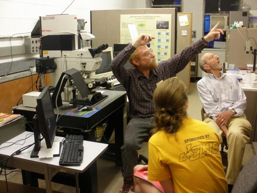
I listened to him discuss his project in further detail and understood everything he was researching and why he needed to alter the microscope for better results. Having experience with fluorescent tagging and confocal/electron microscopes as well as having performed extensive research with off axis illumination, I hope that I could be of help to Dr. Sokolov in some way, shape, or form. Since his project involves lasers, maybe I will be able to work in collaboration with him on a bio/optics project...how exciting!! This was an extremely fantastic day!!
21 June 2011
Returning to my quest to find information on the diffraction theory of a microscope, I spent the morning searching for books to allow me to gain a deeper understanding of the topic. I looked through handbooks and image processing manuals, but found nothing specifically related to what I was interested in. Luckily, I found my favorite optics book in Dr. Noé's office, not recognizing it at first as it is an earlier edition. This helped me greatly in gaining information on diffraction associated with microscopy and reviewing the optics associated with the imaging technique, but I will still need to find associated research.
Pausing my book search, I attended the weekly REU lunch/seminar. This week's seminar included the demonstration of several past REU projects and their associated mathematical proofs as presented by Dr. Graf. He demonstrated the Fermat principle of least time, as I discussed previously in this journal, as well as thermal expansion, reflection, gravitational pull, and parallel plate capacitance. The projects were all interesting and showed a diverse sampling of physics research. Working in the LTC, I focus on optics everyday, but it was nice to break and review other areas of physics for a bit of the time with the other physics REU students.
After lunch, one of my lab mates set up the optical fiber experiment successfully. It was interesting to see the setup. The rest of the afternoon, was simply spent cleaning and organizing the lab.
20 June 2011
Today we derived Young's equation for double slit diffraction. It was a fairly simple process that related several previously discussed topics. Having a particular interest in diffraction, this derivation reminded me of some questions I had concerning near and far field diffraction and the imaging of Airy disks. Luckily, I was able to set up a laser through a pinhole to produce an Airy pattern, altering either the pinhole-to-screen or lens-to-pinhole distance to induce near or far field diffraction. In the far field, the airy pattern was normal - the center of the resulting disk was bright. In contrast, the near field airy pattern produced a disk with a dark center. This experiment was interesting and helped to mend the loose ends of my understanding of the produced patterns. It was fun to actually conduct an experiment in the lab, even though it was fairly simple in nature.
After lunch, I began to research project ideas. I was encouraged by Dr. Marty Cohen to review the diffraction theory of a microscope in hopes of finding research articles combining my individual interests. There was not much available online, so I hope that books are a better resource. After exhausting my internet resources, I went to help a fellow lab member set up a photo-detector to determine the pulse rate of a nitrogen laser. Helping this experiment allowed me to learn much about how the optical tables and accessories work. I also spent time reviewing oscilloscopes and learned about the functions/capabilities of photo-detectors. This project was interesting and informative. It was also fun to collaborate with the other lab members on a project.
16 June 2011 - 17 June 2011
In contrast to yesterday's research, I was actually successful in finding information on different types of resolution enhancing techniques that are not paired with expensive technologies. These types include, but are not limited to, STORM, SSIM, SPEM and OFM. The first three techniques are based on fluorescence microscopy and photon excitation, whereas the last is based on image "scanning." I spent a significant amount of time - by that I mean two full days - attempting to work out the logistics of the optics associated with these techniques and feel that I may be able to use them for my project. Hopefully a lab tour will enlighten me on what sort of optical devices and technologies are available to me, so that I may see what techniques I can use for a project.
The lab tour showed me many of past students' projects, but I did not truly get a sense of what optical devices were available to us. We saw interesting elements such as fiber optic cables and different types of lasers, but I was more interested in looking at the microscopes to get a feel for what I, hopefully, will be working with. I might take another walk around the back of the lab to see if there are any objects related specifically to the research I am reviewing.
15 June 2011
I spent a good amount of lab time today attempting to research resolution in conventional optical microscopes. Unfortunately, I wound up finding paper after paper discussing STED and near field microscopy techniques, as I had wished to avoid. I also attempted to play with the formatting of my web-page, but I gave up because I was not able to find a color combination of text and background that I liked. Maybe tomorrow I will have more luck with both of these subjects and turn out some positive results...
Although I had a most unsuccessful day in lab, I did enjoy the talk given over lunch. The speaker was Rich Migliaccio, founder of East Coast Optical Technologies, who discussed his career path and experience with optics. It was interesting to see the wide range of field experience that he has gained throughout his life, working with different laboratory and technological corporations. He has spent time with various projects, ranging from the development of military intelligence to airport security to tiny bar code scanners to the "Gammacam." Rich has now started his own company which he uses for consulting and developing new technologies. His product, the "Gammacam," is currently helping in the clean up of the nuclear power plants in Japan by imaging gamma particle hot-spots.
I truly enjoyed the lunch seminar. Hoping to one day obtain a career in industry for optical design, the presentation showed me just how interesting the field is and how many paths there are to take. I was intrigued by all of the projects Rich discussed and the purposes that they served. I hope that there are more presentations similar to Rich's, so I may get an even better feel for industry and what it entails. I am extremely glad that I have taken an interest in optics! I feel as if there are barely any limits to what I will be able to work with in the future!
14 June 2011
Today the other REU members and I were taken on a tour of the Van de Graaff building where we learned some of the logistics of the machine and various applications. We were shown the power supplies and vacuum system along with the target room and the area where the linear accelerator used to be housed. Although some of the technical information was over my head, I still found it exciting to tour the area and see the gigantic Van de Graaff machine. It was also interesting to see just how large the S level of the physics building was and the scientific machinery it held. Coming from a small school, I was bewildered by all of the extra space!
The rest of my day was spent researching STED (Stimulated Emission Depletion) and near field microscopy techniques. I looked for some articles and books concerning these techniques in the Science and Engineering library as well, but they were too specific for what I was interested in finding out. Although I find the resolution capabilities of STED and near field microscopy fascinating, I feel that I will not be able to successfully incorporate the techniques into a project, simply due to the lack of the sophisticated and expensive technology that they require. If I begin tinkering in the lab with different optical elements, maybe I will be able to find something interesting to work with that could lead to a feasible project. Reading journal article after journal article is allowing me to research some fascinating topics, but is leading me to become interested in things that cannot be accomplished in this lab alone. I am not sure in which direction I should be headed at this point, but hopefully tinkering in the lab, reading more research articles, and questioning other professors will lead me to an appropriate and interesting project.
13 June 2011
Today we spent much of the morning working on our journals and web pages. I learned a few new html codes to help with formatting so that my journal would not be simply raw text. I have still not gotten the document to appear exactly how I want it, so I will need to learn more codes to correct this issue in the future. The following codes have helped greatly in formatting so far:
h# : Changes the text to header style. The size of the text changes according to the number following the h.
hr : Adds a line rule to the text
head or body: Indicates formatting for the head or body of the text
align="center" width="#" : Increases the "margins" and aligns the text in the center of the page.
The afternoon at the LTC was spent re-capping several topics we had gone over during the previous week, as well as discussing the direction our individual research has taken us. I have recently taken an interest in near field microscopy, but had some questions concerning the technique's ability to accurately image and resolve specimen due to changes in the airy-disk pattern at close ranges. I pondered the fact that, like the pie plate experiment, the image might actually be a shadow of the specimen being viewed, but I feel that this is not an accurate description of the image formation. Hopefully I will be able to achieve a better understanding of this microscopy technique through more research.
Following the discussion of our projects, we reviewed the simple pendulum. We began with a review of the simple forces associated with the pendulum and its direction of motion. Interestingly enough, I determined that small angle approximation could be used to simplify the derivation of the equation of motion. This has given me more insight into just how many topics can be made simple through the incorporation of small angle approximation.
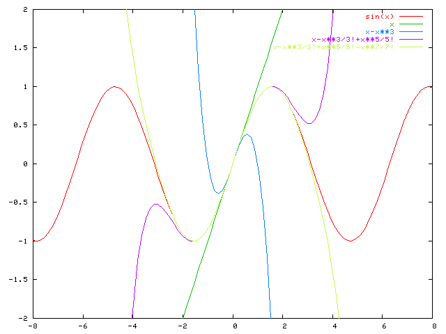
The final topic of the day was a discussion of series expansion, which I have no particular background in. I understood how the equation was constructed and why it was particularly relevant as per small angle approximation previously, but I finally grasped its purpose when a graph was drawn that explained the equation. I now understand that the inclusion of certain terms of the expansion serves the purpose of gaining more and more accurate estimates in small angle approximation. The graphical representation that was drawn on the board and then plotted using the Linux server helped me greatly in understanding the logistics of the equation. The plot is included below:
10 June 2011
The majority of the day was spent researching topics for potential projects and manipulating the web-site. I began the day looking through several OPN magazines to get a feel for different types of optics research in a more informal manner. I believed that by doing this, certain topics would catch my attention more than others. This was a great plan, as it led me directly to research topics in optics with biological applications, which I hope to work with in the future.
I first began researching Raman spectroscopy which I had known to have chemistry applications. In chemistry, Raman spectroscopy is used to view crystalline structures and give information regarding vibrational and rotational modes of specific systems. It can also be used in biology and biochemistry to give information on certain systems such as enzyme/substrate interactions. I set this topic aside as a possible idea for a project, but continued looking for other areas of interest as not to limit myself to the first topic I found.
Another interesting biology-related topic that I ran into in my research was 3-D imaging. I have had some experience with this microscopy technique working with a confocal microscope, but I found that there are other ways to achieve a three dimensional image using microscopy. Specifically, I looked at DHM - digital holographic microscopy - which uses a computer reconstruction algorithm to form an image of the object being viewed. The image is then recorded as a sort of digital hologram. This may be an interesting subject to study or develop a research project on.
The final topic that I came across through the day's research was the construction of a microscope from CD and DVD pickups. The microscope that I read about was capable of fringe projection and 3-D profilometry as well as having fine focus capacity and high resolution. It is interesting that a microscope can be constructed from something as simple and inexpensive as the "guts" of CD and DVD players to create a functionally accurate viewing system! I believe that this would be a great project to work on in the lab. It would also be interesting to alter the conformation of/optical elements involved in the device to allow it to have different applications or be enhanced in certain ways. I have a great interest in developing a project of this nature...This is so cool!
9 June 2011
Our morning at the LTC began with a discussion of html coding language and the Linux server. We learned how to navigate our web page source code and create/edit files. I then went on my own to try out the programming language out on my own to begin documenting my experience and adjusting my web page. I have found that a few commands are helpful in doing this:
cd dir : Changes the current directory to whichever directory is substituted in for "dir."
mv or cp file1 file2 : Moves or copies "file 1" to "file 2."
pico or vi file : Opens file to reveal contents. Text editing can occur here.
more file : Shows the contents of the file in the window. Text editing cannot occur here.
After breaking for lunch, the other researchers and I ventured to the physics library to conduct research on initial project ideas. Since reviewing diffraction and learning about near and far field imaging, I decided to begin searching for project ideas in this area. I first stumbled upon cornu spirals which are a way to graphically document diffraction patterns. These spirals are commonly used to represent diffraction associated with opaque objects or straight-edged objects. In some documents I reviewed, researchers even used cornu spirals to analyze diffraction around objects such as screws!
Researching cornu spirals led me to the topic of fresnel diffraction. The Fresnel equation is often used in near field microscopy, where aperture to screen distance is finite. Other topics associated with this type of diffraction include the Fresnel number, Fresnel integrals, and Fresnel zones. Not having any experience with this topic, explanations of this sort of diffraction seemed tedious and complex. It was suggested to me that the topic was also somewhat obsolete, where most people turn to Fraunhofer or Fourier optics for similar explanations instead. I am not sure if this is the topic I wish to research in, because I feel that I will be frustrated by it. I believe that I will re-start my research on the optics of microscopes, seeing as I am more knowledgeable of the subject and hold a great interest in it. Hopefully this will lead me to better ideas for projects than my search in diffractive optics did.
8 June 2011
Day two at the LTC commenced with a discussion of the outdoor experiments conducted the previous day. After much diagramming and deliberation, we came to several conclusions concerning our data:
- Pie Plate and Flat Lens Experiment
We first documented our results graphically. By moving the pie plates and the flat lens closer and further from the screen where the spot of light was projected, we determined that the size of the spot and the distance to the screen are related in a hyperbolic fashion. Our graph consisted of a hyperbolic function, representing a plot of spot size vs. distance to screen, drawn between the constraints of y=x and y=-x. This validates the observation that while moving the plate closer or further from the screen at close distances, the spot size barely changes, but at greater distances from the screen, the change in spot size is greater.
We then described our results through the use of diagrams and small angle approximations. We found that by using the ratio of the sun's diameter and its distance to the earth and the size of the spot of light created by the pinhole in the pie plate or by the flat lens that the distance from the lens or pinhole to the screen could be determined. This distance was also able to be determined through the use of the equation r=αf, where r is the size of the object, α is ½ the angular size of the sun, and f is the distance from the lens or pinhole to the screen. Using these ratios again in coordination with small angle approximation techniques, the angle of light incident at the center of the pinhole or lens was determined to be 0.01 radians.
Finally, we determined that there was a tradeoff between brightness and image clarity for the pie plate experiment. We found that a smaller pinhole produced a dimmer, yet sharper spot while a larger spot produced the converse.
The conclusions of our experiments led us to the discussion of several other topics...
- Magnification and the 4f System
- Diffraction
We began a discussion of the simple magnification equation and related our experimental data to the equation to learn specific manipulations of variables leading to other relevant equations. We then moved on to discuss the 4f system. To obtain this "system," we created a quadratic formula by plugging a simple distance equation into another relating object and image distances to focal length. From this equation, we determined that if the sum of the object and image distances is less than four times the focal length, no image may be produced.
Overall, it was a challenging, yet informative day.
7 June 2011
Day one at the LTC proved to be both interesting and informative. The other researchers and I learned and reviewed optics through calculation and theory, as well as through hands-on experimentation. We reviewed topics including real/virtual images, conic sections, the Fermat Principle, and small angle approximation. In addition, we discussed series expansion, the Euler equation, and the design of acousto-optic modulators and particle accelerators in short.
Topics discussed in greater depth:- Real and Virtual Images
- Conic Sections
- Fermat Principle of Least Time
- Small Angle Approximation
The later part of the day at the LTC was spent experimenting with lenses, pinholes, and mirrors outdoors. We started by using magnifying lenses to focus light on black paper. The intensity of the light was great enough to burn the paper. Next, we moved on to looking at reading glasses to determine their shape and found that certain lenses were not capable of burning paper due to their focal length to spot diameter ratio. In addition to the reading glasses, we looked at a large, flat lens and two different sized pinholes in pie plates. Holding up the plates and lens so that each spot of light was in focus on a screen, we measured spot diameter and focal length for each object. Interestingly enough, the focal lengths and spot diameters were similar for both pinholes and the flat lens. Finally, we experimented with a flat mirror and determined that if one was to point the mirror directly at the sun, the image produced by the mirror would be exactly the same size as the sun. The results of the other experiments are to be determined tomorrow.