
Background
Current radiation therapy is carried out using mostly high energy x-rays. The current MeV x-rays being used have good qualities such as high tissue penetration and a skin-sparing effect. Despite this, several drawbacks are associated with this method; most importantly, the tissue surrounding the target receives radiation, which is damaging to the tissue. These high energy x-rays interact in a scattering mode rather than a photoelectric one, which would be more confined within the target. To address this issue, a grid theory was developed using orthovoltage x-rays. These techniques would be beneficial if they offered tissue-sparing to healthy tissue near the target tissue; without this quality, it doesn't solve the issue of damaging normal tissue. A need for a method of radiotherapy using orthovoltage x-rays for effectively treating tumors is necessary, but the skin and tissue surrounding the target tissue must be spared in order for this method to be effective.
Technology
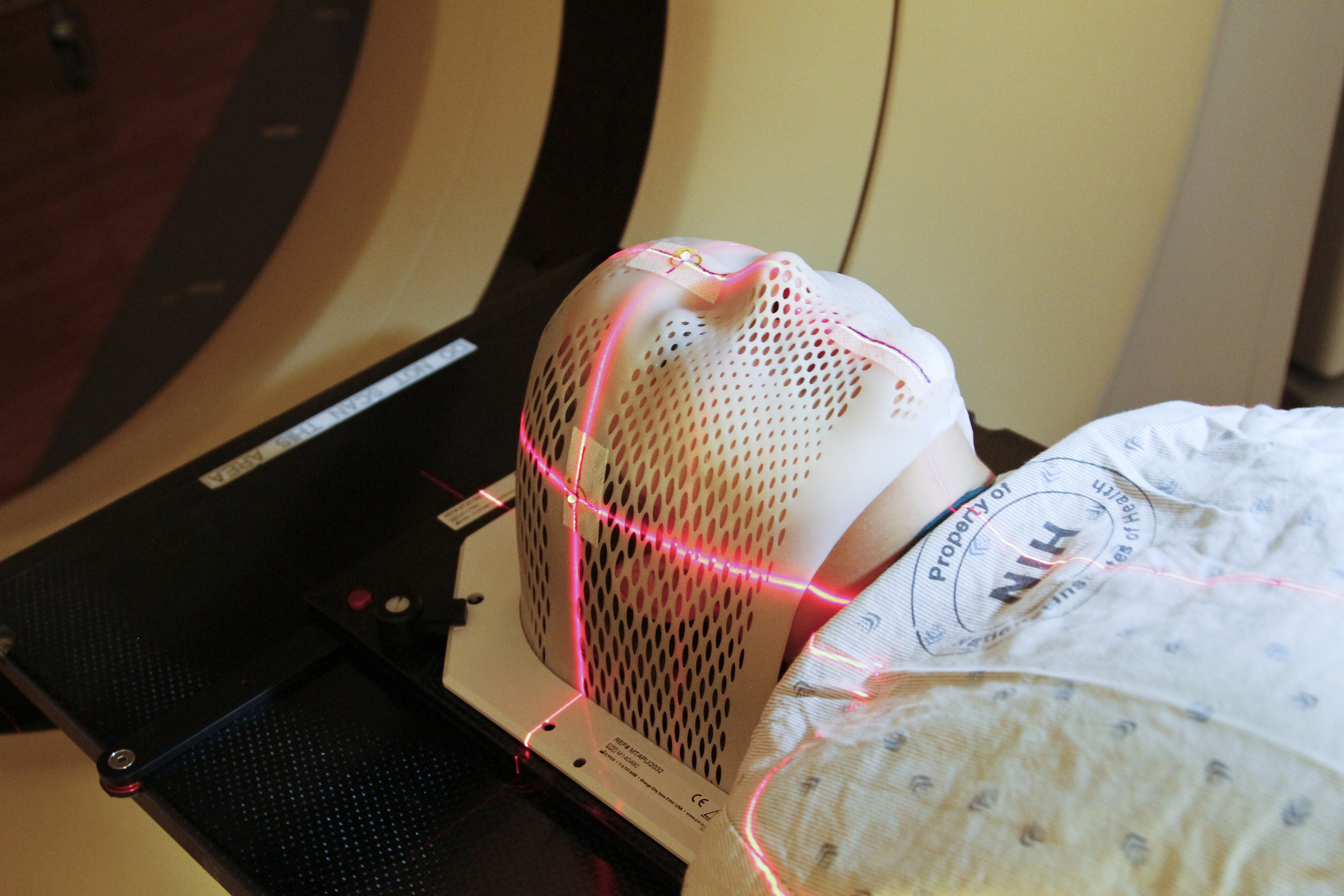
This technology is a system and method for treating a tumor while sparing the surrounding healthy tissue; it uses orthovoltage x-ray radiation. This technology also discloses how to control the tissue depth reached by the radiation, which is essential in order to protect surrounding healthy tissue. This system uses a multi-aperture collimator near the surface of the skin; this lies within a trajectory of radiation, and generates an array of minibeams, which are designed to merge, forming an effective radiation beam.
Advantages
- Low-cost radiotherapy - Compact solution for radiotherapy- Controlled beams of radiation to minimize damage of surrounding tissue - Tissue depth can be varied from around 1 cm to 10 cm - Beam broaden to less than 1.0 mm during merging process - Angular position of the x-rays can be changed to hit target from different angles
Application
- Treatment of tumors - Treatment of neurological targets, such as: - Brain tumors - Disorders, such as epilepsy and alzheimers
Inventors
Avraham Dilmanian, Research Professor, Department of Radiology
Licensing Potential
Development partner,Commercial partner,Seeking investment
Licensing Status
Available for partnership.
Licensing Contact
Donna Tumminello, Assistant Director, Intellectual Property Partners, donna.tumminello@stonybrook.edu, 6316324163
Patent Status
Patented
10124194
Tech Id
8592