
Background
Human review of diagnostic tissue is qualitative, and is therefore prone to high amounts of inter- and intra‑observer variability. Digital pathology, or the review of digitized pathology slides is gaining traction, precisely because quantitative measurements on digitized whole slide images, leads to reproducible and significant nuances observations. The recent approval of whole slide imaging for primary diagnostic uses is leading to widespread adoption of digital whole slide imaging. It is widely expected that within 5-10 years the great majority of new pathology slides will be digitized. Being able to quantitate reliably with reproducibility would be a significant advancement in diagnostic and prognostic applications of digital pathology.
Technology
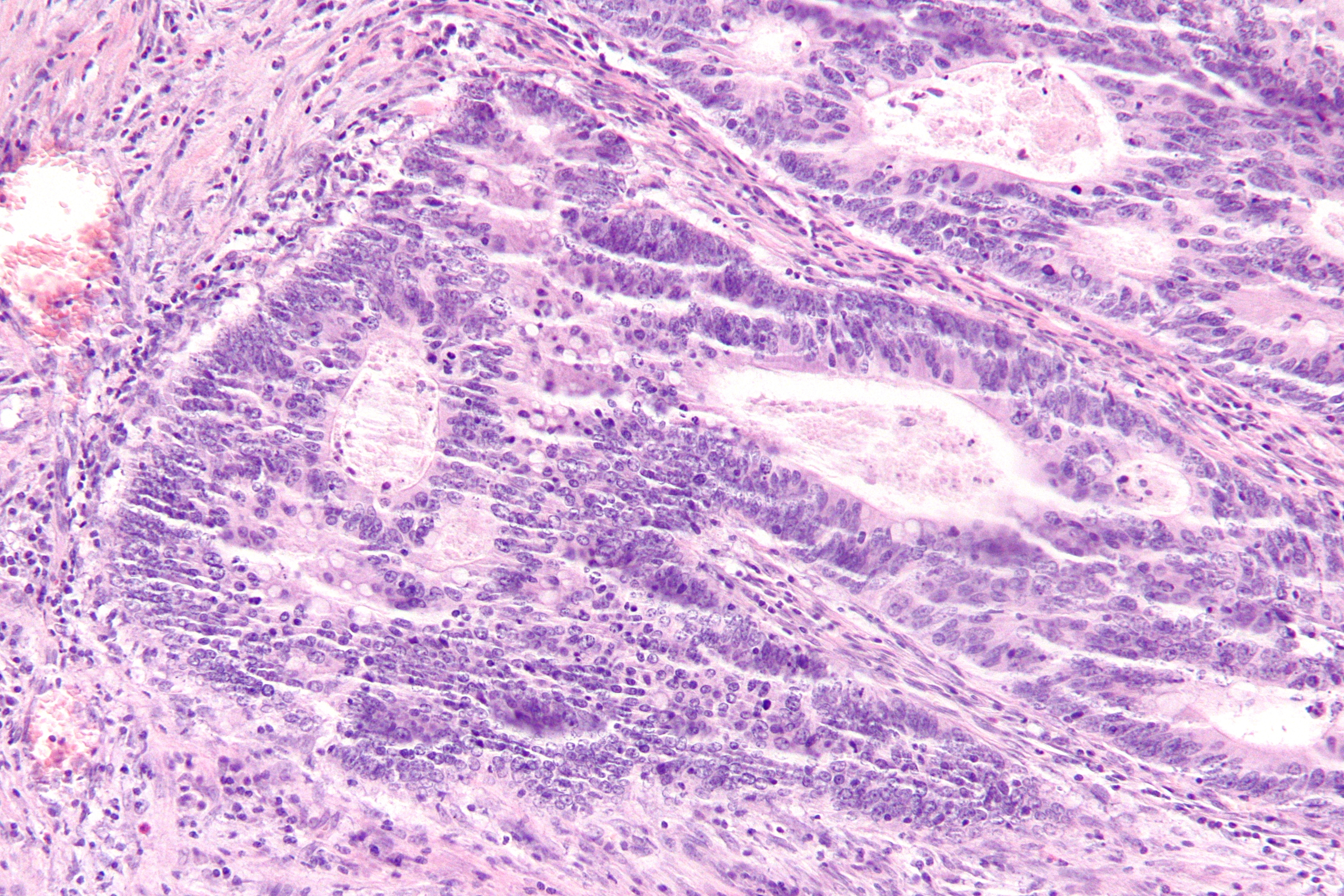
Researchers at Stony Brook University, in collaboration with colleagues at Emory University have developed a novel approach, embodied in methods and software, to extract, quantify, characterize, and correlate Tumor Infiltrating Lymphocytes (TIL) using digitized H&E stained diagnostic tissue slides that are routinely obtained as part of cancer diagnosis. The technology utilizes novel deep learning algorithms to derive a “computational stain”. The computationally stained TILs correlate with the pathologist’s interpretations and with molecular estimates. TIL patterns are linked to tumor and immune molecular features, cancer type and outcomes. This technology provides a robust and reproducible tool for assessment of TILs for diagnostic, prognostic and research purposes.Figure 1 Further Details: Saltz J. et al. 2018 Cell Reports 23, 18-200
Advantages
- High resolution whole slide image analysis of TILs - Scalable - Generalizable, utilizing routine H&E stained slides. - Utilizes low computational power
Application
- Improving diagnostic and prognostic power of digital pathology for assessing TILS - Translation research using digital pathology
Inventors
Joel Haskin Saltz, Chair, Dept. of Biomedical Informatics, Biomedical Informatics
Tahsin Kurc, Associate Professor and Vice Chair, Bidmedical Informatics
Rajarsi Gupta, Clinical Instructor, Biomedical Informatics
Tianhao Zhao, Selective Pathology Fellow, Pathology
Rebecca Batiste, Clinical Instructor, Pathology
Le Hou, Graduate Student, Computer Science
Vu Nguyen, Graduate Student, Computer Science
Dimitrios Samaras, Assistant Professor, Computer Science
Ashish Sharma, PhD - Assistant Professor, Biomedical Informatics Dept.
Vesteinn Thorsson, Senior Research Scientist,
Ilya Shmulevich, Professor,
Arvind Rao, Assistant Professor,
Pankaj Singh, Research Scientist,
Alexander Lazar, Prpofessor,
John Van Arnam, Fellow,
Licensing Potential
Commercial partner,Licensing,University spin out
Licensing Contact
Sean Boykevisch, Director, Intellectual Property Partners, sean.boykevisch@stonybrook.edu, 6316326952
Patent Status
Patent application submitted
PCT/US2018/63231
Tech Id
050-8966